Transforaminal Epidural Steroid Injections

Transforaminal Epidural Steroid Injections

Transforaminal Epidural Steroid Injections

Chronic pain can significantly impact one’s daily life, prompting the exploration of effective interventions. Among these, transforaminal anterior epidural steroid injections have emerged as a valuable non-surgical option since their introduction in 1952. This minimally invasive procedure directly targets the source of pain in the spine, offering relief for conditions such as herniated discs, spinal stenosis, and nerve root compression. In this article, we delve into the key aspects of transforaminal epidural injections, covering indications, contraindications, procedural details, and post-treatment considerations.

What is Transforaminal Epidural Injection?

Transforaminal epidural injection is a procedure in which corticosteroids are injected into the epidural space through the neural foramen, the opening through which the spinal nerve exits the spinal canal. These injections provide a focused solution for individuals grappling with pain stemming from nerve irritation in the neck, back, arms, or legs. By directly administering corticosteroids into the epidural space surrounding the spine cord, healthcare professionals aim to mitigate inflammation associated with conditions like herniated discs, spinal stenosis, and nerve root compression, offering targeted relief where it’s needed most.

Transforaminal Epidural Steroid Injections

Indications for transforaminal epidural injection

Transforaminal epidural injections are commonly used to manage conditions such as lumbar radicular syndrome, which is characterized by pain, numbness, or weakness that radiates along the path of a compressed or inflamed spinal nerve root. It can be caused by conditions such as herniated discs, spinal stenosis, or degenerative disc disease.
These injections are especially recommended for individuals grappling with persistent discomfort in the neck, back, arms, or legs due to conditions such as herniated discs, spinal stenosis, or sciatica. When conventional pain management approaches prove insufficient, pain management specialists may turn to transforaminal epidural injections to directly target and alleviate inflammation at its source. The procedure’s efficacy in mitigating nerve-related pain makes it a valuable intervention for patients seeking relief from the debilitating effects of various spinal disorders.

Contraindications

Contraindications for transforaminal anterior epidural corticosteroid injections are crucial considerations to ensure patient safety and optimize treatment outcomes. While this minimally invasive procedure is generally well-tolerated, certain conditions may preclude its use. Pregnant individuals, due to potential risks to the developing fetus, are advised against this intervention. Active infections and bleeding disorders also pose contraindications, as they heighten the risk of complications. A comprehensive assessment of the patient’s medical history is imperative to identify these contraindications and explore alternative pain management strategies. It is through a meticulous evaluation that healthcare providers can make informed decisions, prioritizing patient well-being and tailoring treatment plans to individual circumstances.

How is it performed?

Pre-procedure preparations

An important element of the pre-procedure phase is the consultation with the healthcare provider. During this consultation, patients can expect a thorough review of their medical history, current medications and a discussion of the potential benefits and risks of the procedure. This exchange of information forms the basis for informed decision-making and lays the groundwork for a personalized and patient-centered approach.

Transforaminal epidural injection is preceded by a series of pre-procedural preparations to ensure a smooth and effective intervention. Patients may receive specific instructions regarding medication adjustments, including temporary discontinuation of blood thinners to minimize the risk of bleeding during the procedure. Fasting for 6-8 hours before the procedure may also be recommended.

Monitoring, local anesthesia and sedation

After being positioned on the procedure table, patients’ vital signs such as blood pressure, oxygen level in the blood and pulse rate are monitored. Local anesthesia is used to make the procedure more comfortable. Some patients may need sedatives.

How is the intervention performed?

Transforaminal epidural injection is a meticulously orchestrated process by skilled pain specialists. The patient is placed prone to facilitate access to the spine and fluoroscopic guidance is used to ensure access to the right site for the procedure.

A thin, specialized needle is carefully inserted through a transforaminal approach into the epidural space, a region surrounding the spinal cord and nerve roots. A contrast medium is then administered and fluoroscopy is used to confirm that the tip of the needle is in the correct location.  The therapeutic mixture, which is a combination of local anesthetic and corticosteroids, or sometimes just a corticosteroid solution, is then injected into the area. Targeted placement of the needle is crucial to effectively intervene in the inflamed area and minimize the impact on the surrounding tissues.

During the procedure phase, patients may experience some mild discomfort associated with the insertion of the needle. However, the discomfort is usually temporary and healthcare providers are skilled at managing any immediate concerns to ensure patient comfort and well-being.

Transforaminal Epidural Steroid Injection intervention

Post procedure care

Following the injection, patients enter the post-procedure care phase, a period of monitoring and initial assessment. The duration of this monitoring can vary but is typically short, allowing healthcare providers to monitor for any immediate side effects and ensure the patient is stable before discharge.

Immediately after the injection, it is not uncommon for patients to experience varying degrees of relief, but it can take several days for the benefits to fully manifest. In the post-procedure period, patients are educated on what to expect and provided with guidelines for self-care. This may include physical activity recommendations, restrictions on certain movements, and the use of ice or heat to manage residual discomfort.

Complications of transforaminal epidural injections

While transforaminal epidural injections are generally safe, there are potential complications, including infection, nerve damage, or allergic reactions to the medications used. Patients are usually advised to report any unusual symptoms following the procedure.

Follow-up and long-term management

After undergoing a transforaminal epidural corticosteroid injection, consistent follow-up is crucial for assessing the procedure’s effectiveness and ensuring sustained pain relief. Patients are typically scheduled for a post-procedural follow-up appointment, during which their healthcare provider will evaluate their response to the injection and address any concerns or questions. This collaborative approach allows for personalized adjustments to the treatment plan, ensuring optimal outcomes. Depending on individual responses, further injections may be recommended to maintain and enhance long-term pain management. Open communication between patients and healthcare providers is paramount, as it facilitates the timely identification and resolution of any emerging issues, contributing to an overall improved quality of life for those seeking relief from chronic pain.

Transforaminal epidural corticosteroid injections offer a targeted and effective approach to managing pain associated with various spinal conditions. With careful consideration of indications, contraindications, and a comprehensive follow-up plan, individuals can reclaim their lives from the grips of chronic pain. Always consult with a healthcare professional to determine the most suitable treatment plan for your specific situation.

Suprascapular Nerve RF Ablation for Chronic Shoulder Pain

Suprascapular nerve radiofrequency ablation for chronic shoulder pain

Suprascapular Nerve Radiofrequency Ablation: An Effective Solution for Chronic Shoulder Pain

 Chronic shoulder pain can have a significant impact on your quality of life, making even simple tasks difficult. Fortunately, there are several pain management techniques available to help alleviate this discomfort. Radiofrequency ablation (RFA) of the suprascapular nerve is a minimally invasive procedure that is gaining popularity as an effective treatment for chronic shoulder pain.

What is suprascapular nerve radiofrequency ablation?

Suprascapular nerve radiofrequency ablation is a minimally invasive intervention to treat chronic shoulder pain by interrupting pain signals from the suprascapular nerve. The suprascapular nerve provides sensory and motor function to the shoulder joint. In cases of chronic shoulder pain, such as osteoarthritis, rotator cuff injury or adhesive capsulitis, the nerve can become a source of persistent discomfort. RF ablation disrupts the nerve’s ability to transmit pain signals, offering relief to patients.

Suprascapular nerve radiofrequency ablation for chronic shoulder pain

Indications for suprascapular nerve RF ablation

Suprascapular nerve radiofrequency ablation is indicated for patients with chronic shoulder pain that has not responded to conservative treatments such as physiotherapy, medications and corticosteroid injections. It is often used to treat conditions such as rotator cuff tears, adhesive capsulitis and osteoarthritis. Other conditions such as bursitis, tendonitis and post-surgical pain that do not respond to conventional treatment may also benefit from this procedure.

Contraindications

Suprascapular nerve radiofrequency ablation is a safe and effective procedure for patients with chronic shoulder pain. However, it is contraindicated in patients with active infection, bleeding disorders, allergy to local anaesthetics, and pregnancy. It should also be avoided in patients who are unable to cooperate during the procedure and in those with severe neurological deficits or weakness in the supraspinatus or infraspinatus muscles, as there is a theoretical risk of weakness in these muscles after the procedure.

How is it performed?

Diagnostic nerve blocks

Before the radiofrequency ablation procedure, diagnostic nerve blocks are performed to confirm that the suprascapular nerve is the source of the patient’s pain. During this procedure, the nerves thought to be causing the pain are temporarily blocked or numbed. This is to determine and confirm that these nerves are responsible for the pain. This relief should only last for about 5-8 hours.

Suprascapular nerve radiofrequency ablation is usually an outpatient procedure performed by an interventional pain specialist. Here is an overview of the procedure:

Preparation

The patient is positioned comfortably and the procedure area is cleaned and sterilised.

Local anaesthetic

A local anaesthetic is administered to numb the area around the injection site, ensuring patient comfort throughout the procedure.

Guided needle placement

Under fluoroscopic or ultrasound guidance, a specialised radiofrequency needle is inserted near the suprascapular nerve. The fluoroscope or ultrasound helps the physician visualise the placement of the needle in real time.

Radiofrequency ablation

Once the needle is correctly positioned, radiofrequency energy is delivered through the needle, generating heat that deactivates the nerve. This disrupts the nerve’s ability to transmit pain signals.

Post-procedure monitoring

After the procedure, patients are monitored for a short period to ensure there are no immediate complications or adverse reactions.

Complications of suprascapular nerve RF ablation

Complications associated with radiofrequency ablation of the suprascapular nerve are rare, but may include minor bleeding, infection, damage to nearby nerves, temporary pain or discomfort, allergic reactions, and weakness in the supraspinatus or infraspinatus muscles after the procedure.

Follow-up after the procedure

Usually, patients are discharged the same day as the procedure and can resume normal activities within a few days. Pain relief may take several weeks to become noticeable, and patients may require additional treatments in the future. Follow-up appointments with the treating physician are recommended to monitor the patient’s progress and ensure that there are no complications.

Suprascapular nerve radiofrequency ablation is a valuable option for patients suffering from chronic shoulder pain when conservative treatments have proven ineffective. This minimally invasive procedure can provide significant relief, improve quality of life and improve overall shoulder function. However, it is important for patients to consult with a healthcare professional to determine if they are a suitable candidate for this procedure and to receive personalised advice about the potential benefits and risks.

Medial Branch Nerve RF Ablation: Solving Back and Neck Pain

Medial branch nerve radiofrequency ablation

Medial Branch Nerve RF Ablation: Solving Back and Neck Pain

Chronic back and neck pain can be debilitating, affecting your daily life and overall well-being. If you’ve tried various treatments with little success, medial branch nerve radiofrequency ablation, also known as medial branch RF thermocoagulation or facet joint denervation, might be the solution you’ve been searching for. In this article, we’ll delve into what this procedure entails, who it’s suitable for, and what you can expect during and after the process.

What is medial branch nerve radiofrequency ablation?

The spine is made up of vertebrae, and between these vertebrae, there are facet joints that provide stability and allow for various types of spinal movements. These facet joints are rich in nerve endings, which are responsible for transmitting pain signals when the joints are irritated or inflamed. The medial branch nerves are small nerve branches that innervate the facet joints, serving as pain messengers.

Medial branch nerve radiofrequency ablation

Radiofrequency denervation is a procedure that uses radiofrequency to desensitize the nerve responsible for the pain, rendering it incapable of sending pain signals on a permanent basis. Medial branch nerve radiofrequency (RF) ablation, is a minimally invasive medical procedure aimed at alleviating chronic pain in the neck and back, primarily caused by issues related to the facet joints in the spine. This procedure specifically targets the medial branch nerves, which play a crucial role in transmitting pain signals from the facet joints to the brain.

Indications for medial branch nerve RF ablation

Medial branch nerve RF ablation is recommended for people with chronic neck or back pain, typically associated with facet joint disorders. Common conditions that may benefit from this procedure include:

Facet joint osteoarthritis: When the joints in the spine become inflamed or degenerate, they can cause severe pain in the back and neck.

Spondylosis: This is a degenerative condition that affects the discs and facet joints of the spine, causing pain and stiffness.

Whiplash injuries: In some cases, patients with unresolved whiplash injuries can develop chronic neck pain that can be treated with this procedure.

Contraindications

Although medial branch nerve RF ablation is effective for many patients, it may not be suitable for everyone. Contraindications include:

Pregnancy: It is not recommended for pregnant women due to potential risks to the unborn child.

Infection at the treatment site: If you have an infection at the site of the procedure, it’s important that it’s treated before you undergo this procedure.

Allergic reactions: Some people may be allergic to the materials used in the procedure.

Bleeding disorders: Patients with bleeding disorders or those taking anticoagulant medications that interfere with blood clotting may not be suitable candidates for medial branch RF thermocoagulation.

Medial branch nerve RF ablation procedure steps

Medial branch nerve blocks (diagnostic phase):

Before undergoing medial branch nerve radiofrequency denervation, patients often undergo a diagnostic phase to confirm the source of their pain. This phase usually consists of the following steps:

Initial assessment: Your healthcare provider will thoroughly review your medical history, perform a physical examination and review any previous imaging studies.

Imaging studies: X-rays, MRI or CT scans may be ordered to help determine the exact location of the source of pain, especially the facet joints in the spine.

Medial branch nerve block: A medial branch nerve block is used to confirm that the pain is coming from the medial branch nerves.

After the diagnostic medial branch nerve blocks, you will be observed for a short period to assess the degree of pain relief. If the diagnostic blocks provide significant relief (more than 50%), you may be considered a suitable candidate for the next stage, which is medial branch radiofrequency denervation.

Medial branch nerve RF ablation procedure

Pre-procedure preparation

Once you’ve been determined to be a candidate for the procedure, your physician will give you pre-procedure instructions. This will usually include instructions on fasting before the procedure, stopping certain medications (such as blood thinners), and arranging transportation to and from the medical facility.

You will have a peripheral vascular line inserted before the procedure and vital signs such as pulse rate, blood oxygen level and arterial blood pressure will be monitored once you are in the procedure room.

Positioning

Before the procedure begins, you’ll be positioned on the procedure table to allow access to the target area. Your healthcare provider will ensure that you are comfortable for the procedure and that you are positioned appropriately in the prone position. Specific positioning may vary depending on whether the medial branch nerve being treated is in the lumbar (lower back) or cervical (neck) region.

Anaesthesia and sedation

After proper positioning, a local anaesthetic is administered to numb the specific area to be treated. This local anaesthetic helps to minimise any pain or discomfort during the procedure. In some cases, conscious sedation may also be used to help you relax and feel more comfortable.
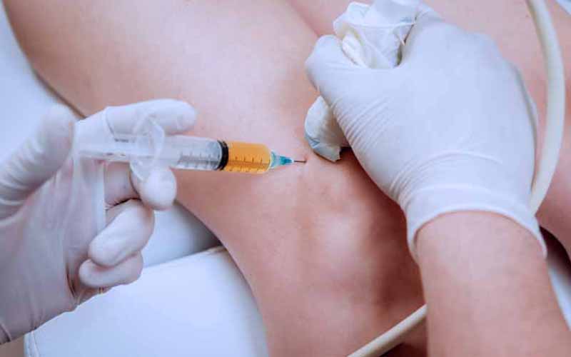
Needle insertion

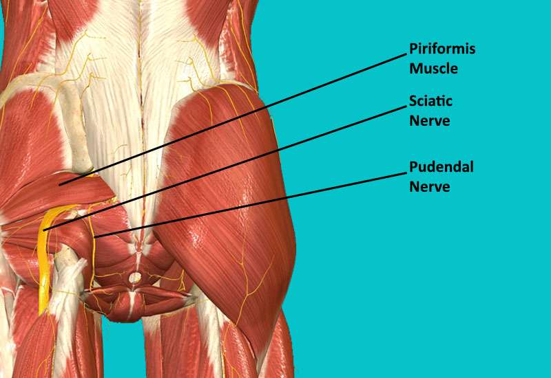
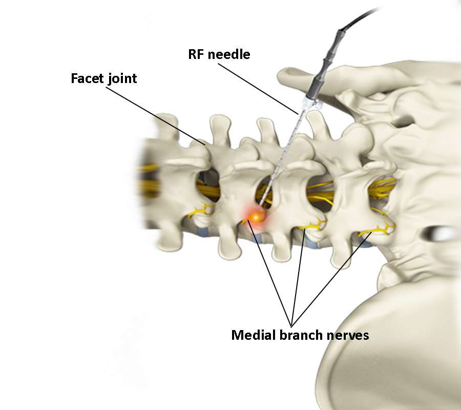
After the anaesthetic and sedation have taken effect, a small, specialised RF needle is carefully guided to the medial branch nerve under fluoroscopic guidance. The aim is to place the needle close to the nerve while avoiding vital structures. After the needle is placed in the right place fluoroscopically, motor and sensory stimuli are given. Small adjustments can be made to be in the closest position to the medial branch nerve.

Medial branch nerve radiofrequency ablation in the procedure room

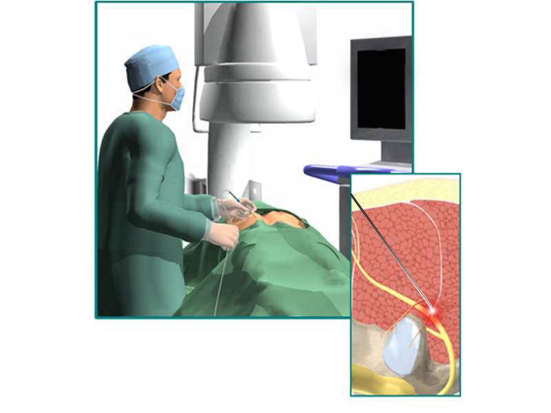
Radiofrequency ablation (denervation or thermocoagulation)

Once the needle is in the correct position, the radiofrequency (RF) ablation procedure begins. RF generates high-frequency electrical currents that heat the tip of the needle. This heat is delivered precisely to the nerve, causing it to coagulate, or essentially ‘burn’, and disrupting its ability to transmit pain signals. The temperatures used are carefully controlled to avoid excessive damage to surrounding tissue.

Post-procedure observation

After the RF thermocoagulation procedure, you will be kept under observation for a short time to monitor your immediate post-procedure condition. This is to ensure that there are no complications and that your vital signs are stable. It’s common for patients to experience mild discomfort or soreness at the treatment site during this time.

Recovery after the procedure

You’ll usually spend a short time in a recovery room where the effects of sedation or anaesthesia wear off. Your healthcare provider will give you post-operative care instructions, which may include recommendations for rest, over-the-counter pain relief, and any activity restrictions. It’s important to follow these instructions to ensure a smooth recovery.

Potential complications of medial branch nerve radiofrequency ablation

Complications associated with medial branch nerve RF ablation are rare, but they can occur. The following are some of the potential complications that can occur with this procedure:

Infection: Infection is a risk common to all invasive procedures, including medial branch nerve denervation.

Bleeding: Bleeding is a rare complication, but is more common in patients with underlying bleeding disorders.

Allergic reaction: A potential allergy is usually to the X-ray contrast agent and rarely to the local anaesthetic.

Aggravation of pain symptoms: In rare cases, patients may experience an increase in pain symptoms after the procedure.

Discomfort at the injection site: Patients may experience some discomfort or soreness at the injection site, but this usually resolves within a few days.

Nerve or spinal cord damage or paralysis: Although very rare, damage can occur as a result of direct trauma from the needle, or secondarily from infection, bleeding causing compression, or injection into an artery causing blockage.

Follow-up and pain relief assessment

In the days and weeks following your procedure, you’ll have a follow-up appointment with your physician. During this visit, your pain relief will be assessed and any necessary adjustments or additional treatments may be discussed.

Keep in mind that the specific details of each procedure may vary depending on the healthcare facility, the healthcare provider’s practices, and individual patient needs. Always consult with your healthcare provider for personalised information and guidance regarding medial branch RF denervation.

Genicular Radiofrequency Ablation for Knee Pain Relief

Genicular nerve radiofrequency ablation -knee pain

Genicular Radiofrequency Ablation for Knee Pain Relief

Knee pain can be debilitating condition, affecting your quality of life and mobility. For those who have exhausted conservative treatments like medications and physical therapy without success, Genicular Radiofrequency Ablation (Genicular RFA) can offer significant relief. In this article, we’ll explore what genicular radiofrequency ablation is, its indications, the procedural steps involved, potential complications, and contraindications.

What is Genicular Radiofrequency Ablation?

Genicular radiofrequency ablation is used to ablate the nerves responsible for transmitting pain signals from the knee joint to the brain. During this minimally invasive procedure, radio waves generated by a radiofrequency device are used to destroy the nerves with the heat generated at the tip of the radiofrequency needle. It is a promising option for those with chronic knee pain that has not responded well to other treatments. Genicular radiofrequency ablation aims to provide long-term pain relief by interrupting these pain signals.

Genicular nerve radiofrequency ablation -knee pain

Indications for Genicular RFA

Individuals meeting the following criteria may be candidates for Genicular RFA:

  • Those suffering from symptomatic knee osteoarthritis (OA) that remains unresponsive to conservative treatments.
  • Individuals with a history of unsuccessful knee replacement surgery, where the exact cause of persistent pain in the region of the total knee arthroplasty remains undetermined.
  • Patients who are not suitable candidates for surgery due to underlying medical conditions and/or a high body mass index.
  • Those who prefer non-surgical alternatives to manage their knee pain.
  • Patients who have previously undergone a successful genicular nerve radiofrequency ablation, as this procedure can be repeated to address recurrent symptomatic knee OA.

Contraindications

GRA may not be suitable for everyone and contraindications include

How is Genicular RFA performed?

GRA is a multi-step procedure that includes preoperative, intraoperative and postoperative phases.

Preoperative phase

A consultation will be conducted by a physician, who will evaluate the individual’s medical history, perform a physical examination, and review relevant imaging studies such as X-rays or MRI scans. Informed consent will be provided to the patient, ensuring they are well-informed about the procedure, its potential risks and benefits, and giving them the opportunity to ask questions.

Intraoperative phase

The patient will be positioned on the procedure table to optimize access to the knee. Medications, such as sedation or local anesthesia, may be administered to ensure the patient’s comfort during the procedure. Imaging techniques such as fluoroscopy or ultrasound will be used by the doctor to accurately identify the target nerves. 

A specialized radiofrequency needle will be inserted through the skin near the genicular nerves. The doctor will assess the pain by stimulating the nerves to determine that the needle is in the right place. Then, with the help of the radiofrequency device, by creating heat at the tip of the inserted needle, a lesion is created in the genicular nerves during a period of 60 – 120 seconds. After the ablation procedure, a mixture of local anesthetic and corticosteroid, usually in a volume of 1-2ml, is injected into the lesioned area.

Postoperative phase

Monitoring patients for a short period is standard procedure to ensure their stability and comfort. In most cases, patients can return home on the same day, but it is essential for them to arrange for someone to provide transportation. Patients may experience some discomfort over the next few days, which should gradually improve. Physicians will schedule follow-up appointments to monitor the patients’ progress.

Complications of Genicular RFA

While the risk of infection at the procedure site is usually very low, however, it is a potential complication that individuals should be aware of. Although rare, there is the possibility of unintentionally damaging nearby nerves during the procedure, which can lead to temporary or permanent complications. Some patients may experience temporary discomfort or swelling at the procedure site, which should gradually subside in the days following the Genicular RFA.

Genicular RFA is a promising treatment option for chronic knee pain in patients who haven’t found relief through conservative methods. It offers a minimally invasive way to target and alleviate knee pain, providing an improved quality of life. However, it’s essential to discuss the procedure, its risks, and benefits thoroughly with your doctor to determine if Genicular RFA is the right choice for you.

Occipital Neuralgia: Symptoms, Diagnosis, and Treatment

Occipital Neuralgia - Nerves

Occipital Neuralgia: Symptoms, Causes, Diagnosis, and Treatment

Occipital neuralgia is a type of headache that causes intense pain in the back of the head, scalp, and neck. It is caused by irritation or injury to the occipital nerves, which run from the top of the spinal cord to the scalp. Occipital neuralgia can be a challenging diagnosis to make, as its symptoms can overlap with other common pain syndromes. In this article, we will discuss the symptoms, causes, diagnosis, and treatment options for occipital neuralgia.

Occipital Neuralgia - Nerves

Symptoms of occipital neuralgia

In typical cases of occipital neuralgia, the most common symptoms of the patient may be the following:

Headache: The most common and prominent symptom of occipital neuralgia is severe, throbbing pain in the back of the head. This pain can come and go or last for a while. The patient may complain of numbness or tingling in that area. Sometimes, certain movements or pressure on the neck can trigger this pain, and dizziness can accompany these symptoms. 

Sensitivity to Touch: The scalp can become hypersensitive to touch. Even light activities such as combing hair or putting the head on a pillow can cause pain.

Eye Pain: This pain can radiate into the eyes and can feel like a headache behind the eyes. Blurred vision, light sensitivity and other vision problems may also occur.

Causes of occipital neuralgia

There are various causes of irritation or damage to the greater and lesser occipital nerves that are considered to be the underlying etiology of occipital neuralgia. Understanding these causes is important in order to manage the condition effectively. Damage to the nerves can be caused by:

Nerve Compression: Occipital nerves can become compressed or trapped by surrounding tissues or muscles. This compression is a common cause of the condition.

Trauma: Head and neck injuries, such as whiplash from car accidents or falls, can damage the occipital nerves, leading to occipital neuralgia.

Tight Muscles: Chronic muscle tension and spasms in the neck and upper back can irritate the occipital nerves.

Medical Conditions: Certain medical conditions like infection, osteoarthritis, diabetes, or gout may increase the risk of occipital neuralgia.

Diagnosis

The symptoms of occipital neuralgia are in some cases similar to those of tension-type headaches and migraines. It can therefore often be misdiagnosed. To correctly diagnose occipital neuralgia, the doctor takes a thorough history and performs a physical examination. Special imaging procedures such as X-rays, computed tomography (CT) or magnetic resonance imaging (MRI) may be ordered to exclude any underlying pathology. When occipital neuralgia is suspected, a diagnostic occipital nerve block may be performed. If pain relief is reported after the nerve block is performed, then the diagnosis of occipital neuralgia is confirmed.

Treatment options

Managing occipital neuralgia often involves a combination of medical and lifestyle interventions:

Physical therapy: stretching and strengthening exercises, hot or cold packs applied to the site of pain usually help patients.

Medication: anti-inflammatory drugs, muscle relaxants, anticonvulsants are prescribed by the doctor if necessary.

Percutaneous nerve blocks: blocks of nerves such as GON and LON, performed by experienced pain specialists, can be used to both diagnose and treat occipital neuralgia

Botulinum toxin (Botox) injections: some studies have reported that Botox injections reduce the pain of occipital neuralgia.

Occipital Nerve Stimulation: this surgical treatment involves placing electrodes under the skin near the occipital nerves. The method works in the same way as spinal cord stimulation and uses the same device. The procedure is minimally invasive and the surrounding nerves and structures are not damaged by the stimulation.

Occipital Neuralgia - Occipital Nerve Stimulation

Spinal Cord Stimulation (DCS): this surgical treatment involves placing stimulating electrodes between the spinal cord and vertebrae. The device generates electrical impulses to block pain messages from the spinal cord to the brain

C2,3 Ganglionectomy: this treatment involves disruption of the second and third cervical sensory dorsal root ganglion.

Lifestyle Modifications: Stress reduction techniques, regular exercise, and maintaining good posture can contribute to symptom management.

Occipital neuralgia can be a challenging condition to manage, but with the right diagnosis and treatment plan, individuals can find relief from the debilitating symptoms. If you suspect you are suffering from occipital neuralgia, it is essential to consult a healthcare professional to discuss your symptoms and explore the most suitable treatment options for your specific case. Effective management can significantly improve your quality of life and help you regain control over your well-being.

Knee Pain: A Comprehensive Guide to Effective Management

knee pain and pain management

Dealing with Knee Pain: A Comprehensive Guide to Effective Management

Knee pain is a widespread issue that can affect people of all ages and backgrounds. Whether you’re dealing with an injury, a medical condition, or the effects of daily wear and tear, knee discomfort can greatly impact your daily life. The good news is that there are various causes and ways to manage knee pain effectively, providing relief and improving your overall quality of life.

What causes knee pain?

Knee pain can have diverse origins, and it’s important to grasp the underlying reasons to effectively manage it. Some common culprits behind knee pain include:

Osteoarthritis: A joint condition that causes cartilage to break down over time.

Ligament injuries: Tears or strains in the ligaments, such as the ACL (anterior cruciate ligament) or PCL (posterior cruciate ligament).

Meniscus tears: Damage to the meniscus, the protective cartilage within the knee.

Tendonitis: Inflammation of the tendons around the knee.

Bursitis: Inflammation of the fluid-filled sacs that cushion the knee joint.

knee pain and pain management


When to seek expert advice?

Recognizing when to consult a medical specialist is crucial when dealing with knee pain. It’s advisable to seek expert guidance if you experience:

  • Severe pain, swelling, or instability in your knee.
  • Difficulty walking or performing daily tasks due to knee discomfort.
  • Persistent knee pain that doesn’t improve with rest or over-the-counter remedies.
  • A history of knee injuries or chronic knee issues.
  • Signs of infection, such as redness, warmth, or fever.

Diagnostic tools for identifying the cause of knee pain

To pinpoint the root cause of knee pain accurately, healthcare professionals may employ various diagnostic tools, including:

X-rays: These reveal bone structure and any signs of arthritis or fractures.

MRI (Magnetic Resonance Imaging): Offers detailed images of soft tissues like ligaments, tendons, and cartilage.

MRI

CT scan (Computed Tomography): Provides a 3D view of the knee joint for precise evaluation.

Ultrasound: Useful for examining soft tissues and identifying issues like fluid buildup or inflammation.

Blood tests: May be conducted to rule out inflammatory or autoimmune conditions contributing to knee pain.

Treatment options for knee pain

When it comes to managing knee pain, a wide range of options is available, catering to various needs and preferences. These options encompass both home-based remedies and professional interventions, ensuring that you can find the most suitable approach for your specific situation.

Home-based remedies

Rest and ice: Begin by giving your knee ample rest and applying ice packs to reduce swelling and discomfort.

Knee pain - ice pack

Elevation: Elevating your leg can help reduce swelling and promote healing.

Over-the-counter (OTC) medications: Non-prescription pain relievers, such as ibuprofen or acetaminophen, can provide temporary relief.

Topical creams: Creams or ointments with anti-inflammatory properties may offer localized pain relief.

Physical therapy

Customized exercise programs: A physical therapist can create a tailored exercise regimen to strengthen the muscles surrounding the knee, improve flexibility, and enhance joint stability.

Manual therapy: Hands-on techniques like massage or manipulation can alleviate pain and improve joint mobility.

Modalities: Therapists may use modalities like ultrasound, electrical stimulation, or hot/cold treatments to manage pain and inflammation.

Medications

Medications play a crucial role in treating knee pain by providing relief from discomfort and managing inflammation. Nonsteroidal anti-inflammatory drugs (NSAIDs) like ibuprofen and naproxen are commonly used to alleviate pain and reduce swelling. Acetaminophen is another option for pain management, although it doesn’t possess the anti-inflammatory properties of NSAIDs. In cases where stronger pain relief is necessary, healthcare providers may prescribe medications tailored to individual needs, considering factors like the severity of pain and the patient’s overall health. It’s essential to use such medications under medical guidance, as they can have side effects and interactions with other drugs. Consulting with a healthcare provider is crucial to determine the most suitable medication regimen for addressing knee pain effectively and ensuring the best possible outcome for the patient’s condition.

Minimal invasive pain management options

Interventional pain management can be a good alternative treatment for patients who are not candidates for prosthetic surgery or who want to avoid more invasive procedures. It is important to talk to your healthcare provider to determine if interventional pain management is a good option for your specific condition.

Corticosteroid injections: These injections can help reduce inflammation and pain in the knee join.

Viscosupplementation: Hyaluronic acid injections can help lubricate the knee joint, reducing friction and pain.

Radiofrequency ablation: A minimally invasive procedure that uses heat to disrupt pain signals from nerves around the knee.

Knee pain - genicular nerve radiofrequency ablation

Platelet-Rich Plasma (PRP) therapy: PRP injections use your own blood components to stimulate healing and reduce pain.

Stem cell injections: This treatment involves injecting stem cells into the knee joint to promote healing and reduce pain.

Braces and supports

Braces and supports can be used to help manage knee pain. Here are some types of braces and supports that can be used:

Knee braces: Knee braces can be used to provide support and stability to the knee joint. They can also help reduce pain and inflammation by unloading the joint. Knee braces can be rigid or flexible, and they can be worn during physical activity or throughout the day.

Knee sleeves: Knee sleeves are a type of knee brace that are made of a stretchy material and provide compression to the knee joint. They can help reduce pain and inflammation and provide support to the knee during physical activity.

knee pain - knee sleeves

Foot/ankle orthoses: Foot/ankle orthoses are medical devices that can be added to a person’s body to support, align, position, immobilize, prevent or correct deformity, assist weak muscles, or improve function. They can be used to decrease pain, improve physical function, and possibly slow disease progression in individuals with osteoarthritis of the knee.

Compression sleeves: Compression sleeves can be used to provide compression to the knee joint, which can help reduce pain and inflammation. They can also help improve blood flow and provide support to the knee during physical activity.

It is important to talk to your healthcare provider to determine if a brace or support is a good option for your specific condition. They can help you choose the right type of brace or support and ensure that it is fitted properly.

Lifestyle adjustments

Weight management: Maintaining a healthy weight can significantly reduce the load on your knees and decrease pain.

Activity modification: Adjusting your daily activities to minimize high-impact movements or using assistive devices when necessary.

Ergonomic changes: Ensuring your work and living environments are knee-friendly, with appropriate seating and support.

Surgical options for knee pain management 

Surgical options for knee pain include:

Knee replacement surgery: This procedure involves resurfacing a knee damaged by arthritis by replacing the damaged parts of the knee joint with an artificial joint.

Knee arthroscopy: This procedure involves inserting a small camera into the knee joint to diagnose and treat problems such as torn meniscus, damaged articular cartilage, and inflamed synovial tissue.

Knee pain - arthroscopy

Cartilage repair: This procedure involves repairing damaged cartilage in the knee joint using various techniques such as microfracture, autologous chondrocyte implantation, and osteochondral autograft transplantation.

Osteotomy: This procedure involves cutting and reshaping the bones in the knee joint to shift weight from the damaged part of the joint to a healthier part.

It is important to talk to your healthcare provider to determine if surgery is a good option for your specific condition. They can help you choose the right type of surgery and ensure that you are fully informed about the risks and benefits of each procedure.

The choice of treatment option will depend on the severity of your knee pain, its underlying cause, and your overall health. Consulting with a healthcare professional is essential to determine the most appropriate approach to manage your knee pain effectively and improve your quality of life.

In conclusion, knee pain is a common issue that can affect anyone, but there are various causes and effective ways to manage it. Consulting with a specialist for an accurate diagnosis and personalized treatment plan can help you regain comfort and mobility in your daily life. By taking proactive steps, you can work towards a healthier, pain-free future for your knees.

Frozen Shoulder and Pain Management Solutions for Relief

Frozen shoulder - adhesive capsulitis

Frozen Shoulder and Pain Management: Effective Solutions for Relief

Frozen shoulder, also known as adhesive capsulitis, can be a distressing condition characterized by pain, stiffness, and limited shoulder mobility. Managing the pain effectively is crucial to improving the patient’s quality of life. In this article, we will delve into the causes and symptoms of frozen shoulder and discuss effective non-invasive and interventional pain management methods to help find relief.

What is frozen shoulder?

Frozen shoulder develops when the capsule surrounding the shoulder joint thickens and tightens, leading to the formation of adhesions or scar tissue. This restricts shoulder mobility, resulting in debilitating pain and stiffness. Although the precise cause of frozen shoulder remains elusive, factors such as diabetes, prior shoulder injuries, and extended periods of shoulder immobility can heighten the risk.

Frozen shoulder - adhesive capsulitis

Symptoms of frozen shoulder

Pain: Persistent pain in the affected shoulder is the cardinal symptom of frozen shoulder. This pain can range from sharp to dull and tends to worsen with movement.

Stiffness: Gradual loss of shoulder mobility is a hallmark of this condition. Simple tasks like reaching for objects or lifting the arm become increasingly challenging.

Limited range of motion: Frozen shoulder typically progresses through distinct stages: the freezing stage (characterized by heightened pain and stiffness), the frozen stage (with persistent pain and limited mobility), and the thawing stage (marked by gradual mobility improvement).

Pain management options

Physical therapy

Physical therapy plays a pivotal role in frozen shoulder treatment. A skilled physical therapist customizes exercises and stretches to enhance shoulder mobility and alleviate pain. These exercises aim to break up adhesions and restore the full range of motion.

Heat and ice

Applying heat or ice pack to the affected shoulder can offer temporary pain relief. Heat relaxes the muscles and increases blood flow, while ice reduces inflammation and numbs the area.

Medications

Healthcare providers may recommend over-the-counter pain relievers like ibuprofen or prescribe stronger medications to manage pain and inflammation.

Interventional pain management of frozen shoulder

In cases where conventional methods may not provide adequate relief, interventional pain management techniques become paramount:

Corticosteroid Injections: These precise injections target the affected area with the guidance of ultrasound, delivering potent anti-inflammatory medication directly to the source of pain.

Shoulder injection

Radiofrequency Ablation: This minimally invasive procedure uses radio waves to disrupt pain signals from the shoulder, providing long-lasting pain relief.

Nerve Blocks: Local anesthetics can be injected near nerves responsible for transmitting pain signals from the shoulder, offering immediate relief.

Hydrodistension

This procedure (hydrodistetion) involves injecting sterile water into the shoulder joint to gently stretch the capsule. It can significantly enhance range of motion and pain relief.

Surgery

In rare cases, surgery may be necessary to treat frozen shoulder. This may involve manipulating the shoulder joint while the patient is under anesthesia, or in some cases, arthroscopic surgery to remove scar tissue or adhesion.

Shoulder arthroscopy - frozen shoulder surgery

Frozen shoulder can significantly impact one’s quality of life, primarily due to the persistent pain and limited mobility it causes. In the journey to manage this condition effectively, interventional pain management techniques have emerged as indispensable tools. Collaborating closely with healthcare professionals, such as pain specialists, can help tailor a personalized pain management plan, incorporating these advanced interventions where necessary. By prioritizing interventional techniques, patients can regain their shoulder mobility and find relief from the often-debilitating pain associated with frozen shoulder.

Regenerative Medicine for Pain: Best Options

trombositten zengin plazma tedavisi

What is Regenerative Medicine? Which Regenerative Medicine Options are Recommended for Pain Treatment?

What is regenerative medicine?

Regenerative medicine is an emerging branch of modern medicine that refers to the process of replacing or regenerating cells, tissues or organs to fulfil their normal function. In the practice of pain management, 3 specific treatment modalities are mainly used as regenerative medicine: platelet-rich plasma (PRP) therapy, stem cell therapy and prolotherapy. Regenerative medicine treatment modalities focus on healing damage to the body and are thought to result in pain relief as well as improved function.

Rejeneratif Tıp - Regenerative Medicine

Now I would like to give you brief information about PRP, stem cell therapy and prolotherapy:

Platelet-rich plasma therapy (PRP)

The patient’s own blood is used for platelet-rich plasma or PRP treatment. Using the patient’s own blood eliminates the risk of infectious disease transmission during treatment and is considered a minimally invasive type of treatment. Blood is drawn from the patient and then placed in a centrifuge. Platelets are separated from part of the blood and then injected into the treated area. Concentrated platelets have high doses of bioactive proteins, such as growth factors, which are essential in the repair and regeneration of tissues.  Platelet-rich plasma therapy injections have five to ten times the amount of platelets normally found in the blood. The quality of PRP depends on the viability of platelets. PRP should be prepared and administered by a specialised team.

PRP - platelet rich plasma

Stem cell therapy

Stem cells are cells that have the capacity to transform into any specific cell type of the body, and are therefore smart cells that can transform into the required cell type according to the signal they receive. Their task is to renew or repair the living structure (e.g. skin, tissue, organs, etc.) of which they are a part. Bone marrow and adipose tissue-derived stem cells are the most commonly used especially in the treatment of chronic pain caused by injuries in the musculoskeletal system. Injecting stem cells into a damaged area of the body is thought to help regenerate healthy tissue.

Kök Hücre tedavisi - Stem Cell Therapy

Prolotherapy

Prolotherapy involves a series of injections of a mildly irritating agent into the area of pain or instability, performed at intervals of several weeks. Classical prolotherapy involves injecting a hypetonic dextrose solution into a specific area where damage or injury has occurred. Although some study results have been published in the medical literature regarding the effectiveness of prolotherapy in the treatment of musculoskeletal pain, high-quality randomised controlled trials supporting these results are limited.

Cancer Pain: Effective Strategies for Management

Cancer pain - kanser ağrıları

Understanding Cancer Pain: Effective Strategies for Management

What is cancer pain?

Cancer is a universal health problem of modern life, and can cause pain as it grows by destroying or pressing on nearby structures in the body, such as nerves, organs and bones. Cancer that has spread from its original site (primary tumour) can move to other parts of the body (metastasis), where it can also damage nearby structures and cause pain. The pain symptoms experienced by patients with cancer can vary from person to person and can range from mild to severe chronic pain.

Cancer pain - kanser ağrıları

Pain can be caused by the cancer itself or by the cancer treatments used to fight it, such as chemotherapy, radiation therapy and surgery. Even today, when different treatment modalities are available, 46% of patients do not receive adequate pain management at the time of death. For this reason, the World Health Organisation has defined cancer pain as a personal right and emphasised that this pain must be relieved.

Is it possible to treat cancer pain?

More than 80% of cancer pain can be controlled by pain management specialists. The aim of pain treatment in cancer is to prolong the patient’s pain-free sleep time, to ensure pain relief at rest, and a pain free life while standing or moving. By providing these, it is to contribute to the patient’s active and quality life as much as possible.

Apart from the physical characteristics of cancer pain, psychological and social consequences of cancer pain also affect the patient’s quality of life. Therefore, it is important to control psychological and social effects during the treatment phase. Evaluation and re-evaluation are of great importance in the treatment of cancer pain. This is important both for monitoring the effectiveness or inadequacy of the treatment and for the recognition of pain of different localisation and character as the disease progresses. Painkillers, interventional pain management procedures, cognitive and behavioural approaches can be used in the treatment.

There are also some barriers to an appropriate approach to cancer pain. The most important of these is the fear of drug addiction in the patient. This concern manifests itself in the form of confusion of psychological and physical addiction, ignorance about drug tolerance and exaggerated expectation of side effects by patients and their relatives. Insufficient prophylactic and therapeutic approaches to an opioid side effect such as constipation, for which tolerance does not develop, also constitute an obstacle to appropriate analgesic treatment approaches in cancer patients.

When determining the treatment method, it is essential to make the decision together with the patient and his/her family, to ensure the patient’s active participation in the treatment, and to inform the patient about side effects. Obtaining the opinions of the relevant departments about pain is of great importance in the evaluation before the treatment plan.

The stepwise pain treatment recommendations of the World Health Organization in 1986 are accepted worldwide and can be modified according to minor differences in approach from centre to centre and from country to country. Chemotherapy, radiotherapy and surgical methods used for the treatment of cancer also contribute to the reduction of pain with the tumour-shrinking effects they provide.

In conclusion, there are many different approaches to improve the patient’s quality of life in the treatment of cancer pain. In these patients, the most effective treatment approach should be planned with the participation of the patient, the patient’s relatives and the physician, taking into account the location, intensity and character of the pain, the type of cancer and the psychosocial status of the patient.

What Is A Migraine? Current Approaches In The Treatment

Migren baş ağrısı tanı ve tedavi

What Is A Migraine? Current Approaches In The Treatment Of Migraine

What is a migraine?

Migraine is a disease as old as human history. This type of headache is a very important health problem for the society. It is a type of headache that tends to recur and causes moderate to very severe pain. The pain is usually described as throbbing or pulsating and usually begins on one side of the head. Migraine headaches get worse with physical activity, light, sound, or physical movement.

what is a migraine?


If left untreated, a migraine headache attack typically lasts from 4 hours to 3 days. Patients may be sensitive to light, sound and even smell and may also experience nausea and/or vomiting. Some of the patients may feel that the pain will come 5-60 minutes before the presence of warning signals known as aura from pain attacks. Migraine is not a life-threatening brain disease. After the diagnosis of migraine, the treatment of the patient should be done according to a certain order and in cooperation with the physician.

Current approaches in the treatment

In migraine attacks, resting in a dark and quiet room with an ice pack can help reduce the pain. If sleep is possible, the patient usually wakes up free of pain. For the prophylaxis of attacks, the frequency of pain can be reduced by some simple measures such as avoiding the factors that trigger the disease, not skipping meals, and ensuring regular sleep times.

Medications for migraine treatment

Medications in migraine is given in two ways as “prophylactic” or preventive treatment to prevent attacks, and “attack (acute headache) treatment” to relieve symptoms of an attack such as pain, nausea and vomiting. Prophylactic treatment is aimed at reducing the frequency and severity of pain by using regular medications for a certain period of time. Migraine treatment is a form of treatment in which the same treatment is not applied to everyone. And each patient diagnosed with migraine should be given a special decision.

Anti-CGRP monoclonal antibodies (erenumab, galcanezumab, fremanezumab), popularly known as migraine vaccine, are approved for use in the preventive treatment of migraine. Although it is called a ‘vaccine’ because it is an antibody, it does not make it completely protected from the disease once it is made, like the vaccines we know in the classical sense. In fact, it is more accurate to define it as a medicine, not a vaccine.

Interventional treatments

Botulinum neurotoxin is used in chronic migraine prophylaxis. Although there is not enough evidence showing its effectiveness in the treatment of episodic migraine, it can be recommended in some clinics. Local anesthetics and greater occipital nerve (GON), lesser occipital nerve (LON), supraorbital nerve (SON) blockade, and sphenopalatine ganglion blockade are among the safe and effective interventional treatments in migraine with frequent attacks, especially chronic migraine.

Botox injection for migraine headache

Neuromodulation

Different neuromodulation (stimulation of the nervous system) methods can be used in the treatment of migraine. There is experience in the treatment of migraine with both invasive and non-invasive methods. Non-invasive methods include transcranial magnetic stimulation, transcranial direct current stimulation, and non-invasive vagal nerve stimulation (VSS); invasive methods include occipital nerve stimulation and implanted VSS. It has no routine clinical use yet.

What is a migraine headache diary?

A migraine headache diary is a written or digital log where individuals can record detailed information about their migraine episodes. It serves as a personal record that helps track and monitor various aspects of migraines, including the frequency, duration, intensity of pain, triggers, and associated symptoms. The diary may also include information about medication usage, lifestyle factors, sleep patterns, and any remedies or interventions tried during a migraine attack.

By consistently maintaining a migraine headache diary, individuals can identify patterns and potential triggers, recognize early warning signs, and track the effectiveness of different treatments or lifestyle modifications. The diary acts as a useful tool for both individuals and healthcare professionals, providing valuable information for accurate diagnosis, personalized treatment plans, and improved management of migraines.

If you’re looking to better understand your migraine headache condition and track your symptoms, “The Migraine Detective: A 30-Day Diary For Solving The Mystery Of Your Headaches” is an affordable option designed by Tural Bayramov, MD.